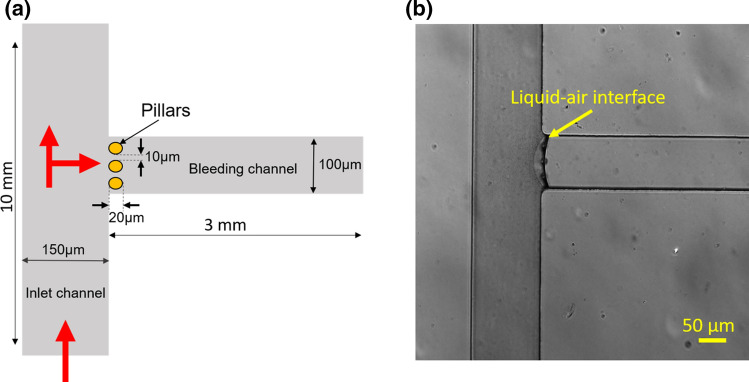
Figure 2.
Design and development of a polydimethylsiloxane based microfluidic bleed chip. (a) The bleed chip consists of an inlet channel of width 150 μm, length 10 mm and a side channel of width 100 μm, length 3 mm referred to as the bleeding channel. At the intersection of channels, there are 3 pillars of 20 μm diameter with a 10 μm gap between them. Red arrows depict the direction of blood flow in the device (Figure not to scale). (b) Differential interference contrast (DIC) 10× image of the device where the bleeding channel is coated with fibrillar collagen (100 μg mL−1). The arrow indicates the liquid-air interface during coating of the bleeding channel. Scale: 50 μm

