Figure 2.
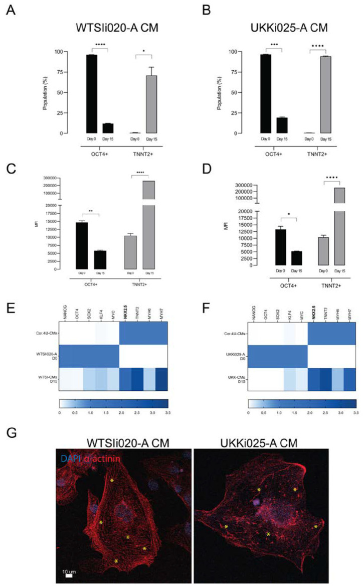
Characterization of cardiomyocytes derived from WTSIi020-A and UKKi025-A. (A,B) Flow cytometry analysis of cells positive for TNNT2 and OCT4 at the beginning (day 0) and the end (day 15) of differentiation in CDM3. (C,D) Median fluorescence intensities of TNNT2+ and OCT4+ populations at day 0 and 15 of the CDM3 protocol. n = 3 biological replicates. (E,F) Heatmap plots representing RT-qPCR for markers of pluripotency (NANOG, OCT4, SOX2, KLF4, MYC), cardiac progenitors (NKX2.5) and cardiomyocytes (TNNT2, MHY6, MHY7) on day 0 and day 15. n = 3 independent biological replicates performed in technical triplicates. (G) Immunofluorescent staining images of cardiomyocytes derived from WTSIi020-A and UKKi025-A. WTSIi020-A CMs show clear formation of immature sarcomeric-like structure while UKKi025-A CMs display a disrupted structure and rather enlarged cell morphology (marked with *). Significance level A: **** poct4 < 0.001; * ptnnt2 < 0.023; B: *** poct4 = 0.0002; **** ptnnt2 < 0.0001; C: ** poct4 = 0.0015; **** ptnnt2 < 0.0001; D: * poct4 = 0.0142; **** ptnnt2 < 0.0001; E: p ≤ 0.0001; F: p ≤ 0.0002.

