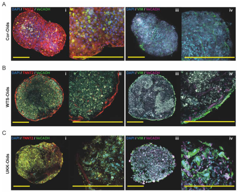
Figure 4.
Determining the structural development of cardiac organoids. (A–C) Immunofluorescence images of Cor-Oids, WTS-Oids and UKK-Oids after 21 days of culture, labelled with cardiac-specific troponin (TNNT2), vascular cadherin (VeCADH) and vimentin (VIM). We identified similar structural patterns (A–C, iii–iv) in all organoid types, in terms of HCMEC (magenta) and HCF (green) distribution. Different structural development patterns were observed regarding the migration of hiPSC-CMs and the formation of cardiac filament-like structures (red), which were well-defined in Cor-Oids (A, i–ii), mostly perimetric in WTS-Oids (B, i–ii), and still in formation in UKK-Oids (C, i–ii). Scale bars = 200 µm.

