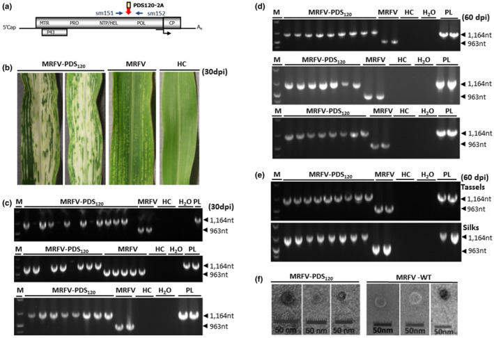
FIGURE 2.

Viability of MRFV HEL/POL junction as insertion site. (a) Location of primers (sm151 and sm152) used for RT‐PCR detection of MRFV‐PDS120 and MRFV‐WT. The primers straddle the HEL/POL junction and amplify 1,164 bp in MRFV‐PDS120 and 963bp in wild‐type MRFV. (b) Virus symptoms and the chlorophyll photobleaching phenotype induced by MRFV‐PDS120 at 30 dpi, compared to leaves of plants inoculated with MRFV without insert and noninoculated leaves (HC). (c) RT‐PCR analysis of virus accumulation in systemic leaves at 30 days post‐inoculation. The three gels show RT‐PCR detection of MRFV‐PDS120 and MRFV‐WT in inoculated plants in three replicated experiments, with RNA from noninoculated plants (HC) and water (H2O) included as negative control templates; and the MRFV‐PDS120 plasmid (PL) serving as a positive control. Blank lanes in MRFV‐PDS120 inoculations represent nonsymptomatic plants. M: 100 bp DNA marker. (d) RT‐PCR analysis of insert retention in MRFV‐PDS120 at 60 days post‐inoculation. RT‐PCR was repeated at 60 dpi only for RT‐PCR positive (symptomatic/successfully inoculated) MRFV‐PDS120 plants in (c) above, with controls and DNA marker included as described above. This was done for all the three replicated experiments. (e) RT‐PCR assays for virus accumulation in tassels and silks for a subset of MRFV‐PDS120 symptomatic plants. Controls and DNA marker were included as described above. (f) Transmission electron micrographs of MRFV‐PDS120 (left panel) and MRFV‐WT particles (right panel) in maize (Silver Queen) extract
