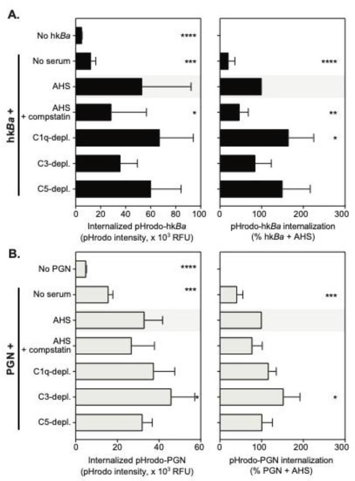
Figure 3.
Quantitation of pHrodo-hkBa (A) and/or pHrodo-PGN (B) internalization by neutrophils in the presence of compstatin, a C3 convertase inhibitor, or the immunodepletion of complement factors. Data are shown as mean ± SD of 6 independent donors, and depict pHrodo fluorescence intensity after the internalization of labeled bacteria or PGN (left panels) and normalized changes in endocytosed pHrodo-labeled particles compared to bacteria or PGN uptake in the presence of autologous serum (AHS), which was considered 100% (right panels). Statistically significant differences compared to pHodo uptake in the autologous serum (AHS, shaded) are depicted graphically (* p < 0.05; ** p < 0.01; *** p < 0.001; **** p < 0.0001), and were computed by repeated measures one-way ANOVA with Holm–Sidak’s multiple comparisons test (left panels), or one sample t test compared to the normalized value (right panels).

