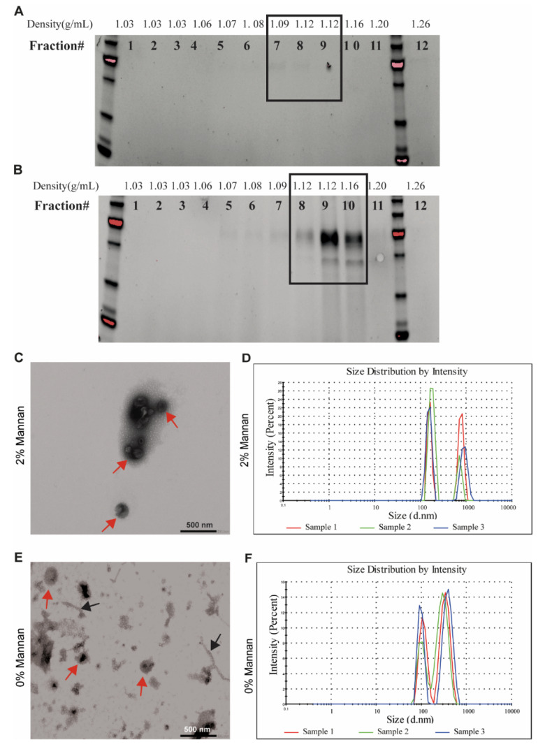
Figure 2.
Characterization of secreted EVs from a pig fecal inoculum. The fecal inoculum was cultivated in vitro either in the presence of 2% β-mannan or without any carbohydrate source for 24 h before removing the cells from the culture medium. Density of EVs isolated from fecal culture on basal medium (A) or supplemented with 2% β-mannan (B) using a density gradient. Twelve fractions were recollected from the top of the gradient and analyzed using SDS-PAGE. The electron microphotograph shows that the sample recovered after ultracentrifugation and density gradient separation was bacteria-free and consisted of vesicles shaped as spheres in the presence of 2% β-mannan (C) or without carbohydrate (E). The red arrow indicates EVs, while black arrows indicate “pili-like” structures. The scale bar indicates 500 nm. Size of the vesicles isolated in the presence of 2% β-mannan (D) or without carbohydrate (F) was visualized by a Zetasizer uV.

