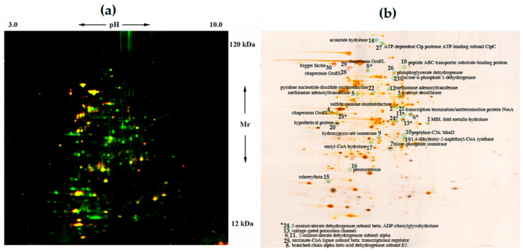
Figure 1.
Two-dimensional (2D) DIGE analysis of Sb. thermotolerans cells grown in the presence of the arsenopyrite concentrate (Cy3 dye, green) and in the control medium (Cy5 dye, red) (a) and a corresponding silver-stained gel (b). Arrows (a) indicate pH (3–10) and Mr (12–120 kDa) ranges. Circled spots (b) correspond to proteins ≥2-fold up- (green circles) or downregulated (red circles) in response to the concentrate (spot numbers refer to Table 3) and identified by MALDI (Matrix-Assisted Laser Desorption/Ionization) mass spectrometry.

