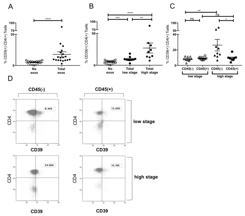
Figure 5.
Induction of CD39+ Treg differentiation by plasma-derived exosomes. (A) Expression of CD39 after incubation with total exosome fraction of patients of all stages (B) Expression of CD39 after incubation with total exosomes of low stage vs. high stage patients. (C) Expression of CD39 after incubation with CD45(−) and CD45(+) exosomes of low stage and high stage patients. Note that only incubation with CD45(−) exosomes showed significant stage-dependent differences.; n = 18. p values were determined with Mann-Whitney test, with * p < 0.05, ** p < 0.005, **** p < 0.0001. Bars represent standard error of mean (SEM). (D) Representative flow cytometry plots depicting CD39 expression of CD4+ T cells after incubation with CD45(−) (left) or CD45(+) (right) exosomes of UICC low stage (top) or high stage (bottom) patients.

