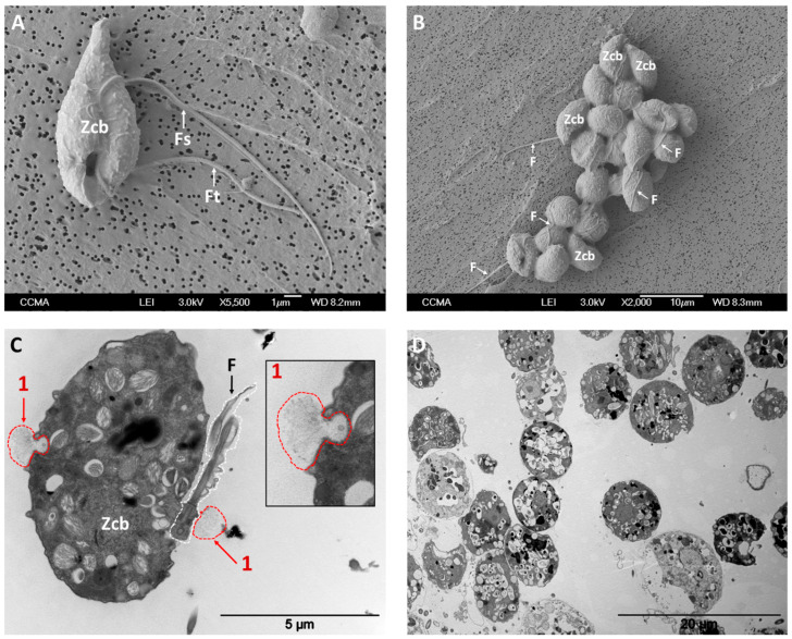
Figure 1.
Micrographs of P. parasitica zoospores at unicellular stage, taken in absence of K+ (panels A and C) and of cell aggregates, taken at t = 15 min after K+ application (panels B and D). Zoospore cell bodies and flagella are indicated with “Zcb” and “F”, respectively. “Fs” and “Ft” indicate smooth and tinsel flagella, respectively. Panel C shows two secretory events (1); the inlet depicts a secretory vesicle that has just secreted its fibrillary content. Panel B shows the presence of several and representative ellipsoidal cells (such as those indicated as “Zcb”) and interwoven flagella (F), indicating a prominent proportion of cells at the zoospore stage in aggregates. Images were obtained through scanning electron microscopy (SEM; panels A and B) and transmission electron microscopy (TEM) on analyses of 80-nm-thin sections (panels C and D) in presence or absence of K+.

