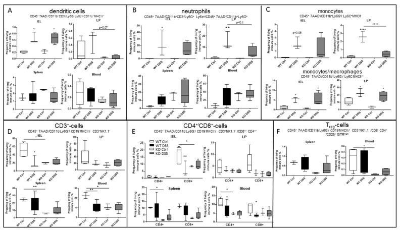
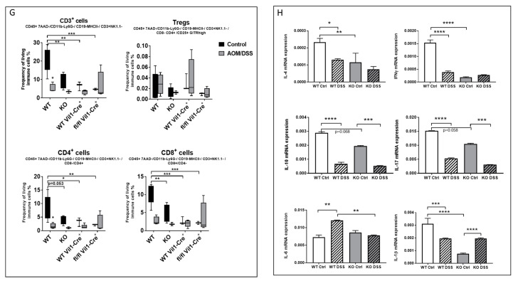
Figure 5.
Flow cytometry analysis of different mice tissues (intraepithelial lymphocytes (IEL), lamina propria (LP), spleen, and blood) upon acute DSS treatment in CerS5-wt and -ko mice. Dendritic cells (A), neutrophils (B), and monocytes/macrophages (C) were increased in the colon tissue fractions after DSS treatment. By contrast, CD3+ T-cells were decreased in CerS5-wt mice after DSS treatment or were already low in CerS5-ko mice (control and DSS-treated) (D–F). Shown are mean ± min/max IEL: WT Ctrl n = 5, WT DSS n = 3, KO Ctrl n = 3, KO DSS n = 4; LP: WT Ctrl n = 5, WT DSS n = 3, KO Ctrl n = 2, KO DSS n = 4; spleen: WT Ctrl n = 3, WT DSS n = 6, KO Ctrl n = 3, KO DSS 6; blood: WT Ctrl n = 3, WT DSS n = 7, KO Ctrl n = 3, KO DSS n = 6. (G) Blood T-cell status in CerS5-wt and CerS5-ko mice as well as in CerS5wt-VilCre and CerS5fl/fl-VilCre mice in the chronic AOM/DSS mouse model. Blood leukocytes were isolated and quantified by flow cytometry. Here, only CD3+-cells are shown. Analysis was performed using FlowJo software v10. Data are mean ± SEM of n = 2–4 animals in each group. (H) Cytokine mRNA expression in colon tissue of CerS5-ko and CerS5-wt mice. RNA from colon tissue was extracted and mRNA of the various cytokines detected by real time PCR. Data are mean ± SEM of n = 4–6 animals in each group. Statistical analysis was performed by one-way ANOVA. p * = 0.05, p ** = 0.01, p *** = 0.001, p **** = 0.0001.


