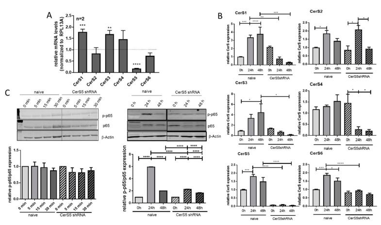
Figure 6.
Activation of Jurkat cells is prohibited by CerS5 downregulation. (A) Transduction of CerS5-shRNA into Jurkat cells led to a significant reduction of CerS5 mRNA expression. (B) CerS mRNA expression was increased after stimulation of Jurkat cells with IL-2 (200 Units/mL) and anti-CD2/3/28 activation beads (1:1 beads-to-cell ratio) for 24–48 h. This was inhibited by CerS5 downregulation. (C) NF-κB activation in Jurkat cells after treatment with IL-2 (200 units/mL) and anti-CD2/3/28 activation beads (1:1 beads-to-cell ratio) for 0–48 h. NF-κB activation was detected by anti-phospho-p65 and anti-p65 antibodies by Western blot method. Data from (A) and (B) are mean ± SEM of 2–4 independent experiments measured in triplicate. (C) One representative blot is shown of three to five. For calculation, the expression levels of p-p65 and p65 were related to β-actin and subsequently related to untreated control cells. Data are mean ± SEM. Statistical analysis was performed by one-way ANOVA. p * = 0.05, p ** = 0.01, p *** = 0.001, p **** = 0.0001. The whole western blot image please find in Figure S4.

