
Figure 3. Sel1L deficiency leads to the formation of megamitochondria with perforating ER tubules.
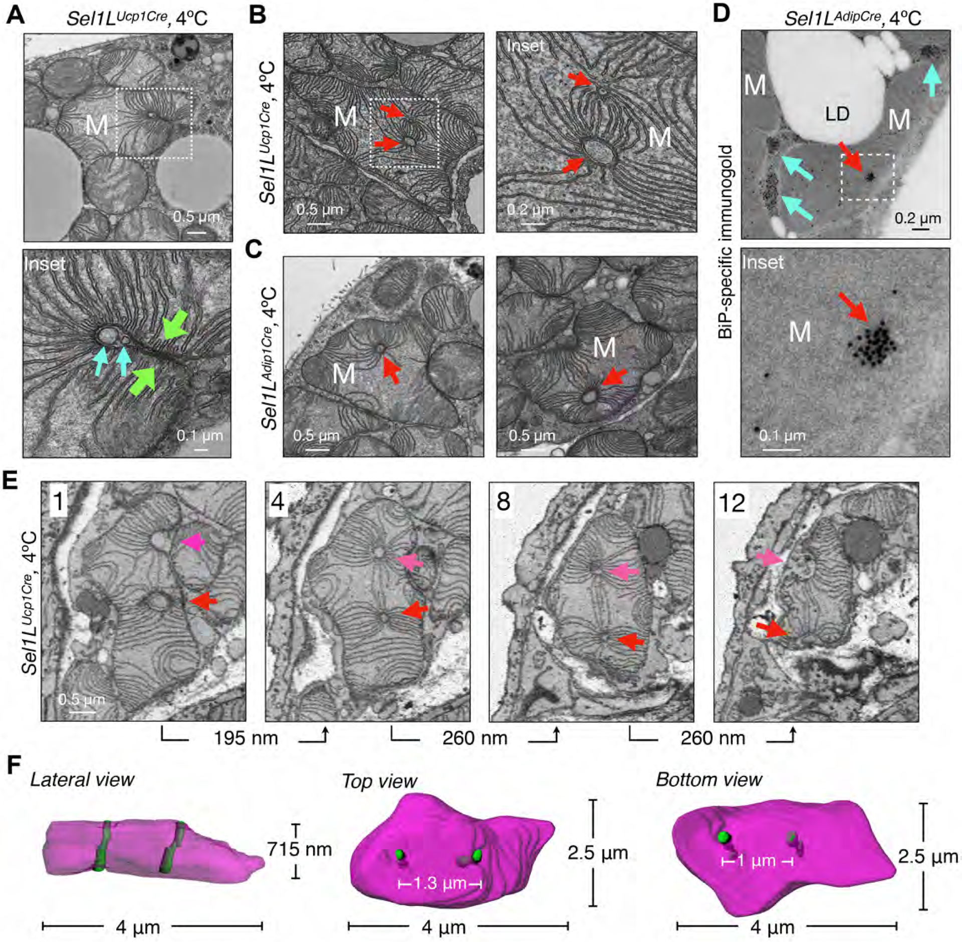
(A) Representative TEM images of BAT in Sel1LUcp1Cre mice housed at 4°C for 6 hr, showing a megamitochondrion wrapping around the tubular structures (cyan arrows). Green arrows, two opposite sides of a mitochondrion. (B-C) Representative TEM images of BAT from Sel1LUcp1Cre (B) and Sel1LAdipCre (C) mice at 4°C for 6 hr, showing megamitochondria with tubular structures (red arrows). (D) Representative BiP-immunogold TEM images of BAT in Sel1LAdipCre mice at 4°C for 6 hr. Red and cyan arrows, mitochondria-perforating ER tubules and peri-mitochondria ER tubules. (E-F) Representative SBF-SEM images (E) and 3D reconstruction (F) in BAT of Sel1LUcp1Cre mice at 4°C for 6 hr, showing four different slices of a megamitochondrion with two parallel perforating ER tubules (red and magenta arrows). All 12 slices (65 nm/slice) are shown in Fig. S8. All experiments have been repeated two to three times.