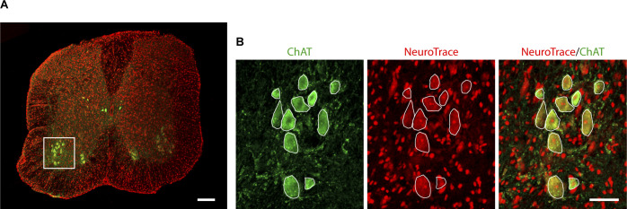
Figure S5. Nissl substance intensively marks ChAT-positive motor neurons.
Nissl substance intensively marks ChAT-positive motor neurons. (A) Representative image of a double labeling for NeuroTrace (red-fluorescent Nissl) and ChAT (green) in spinal cord section containing the lumbar region L1-L5 from 30-dpp WT mouse. The white square highlights the ventral horn of the spinal cord analyzed. Bar, 200 μm. (B) Higher magnification of ChAT, NeuroTrace and merge signals. Bar, 20 μm.

