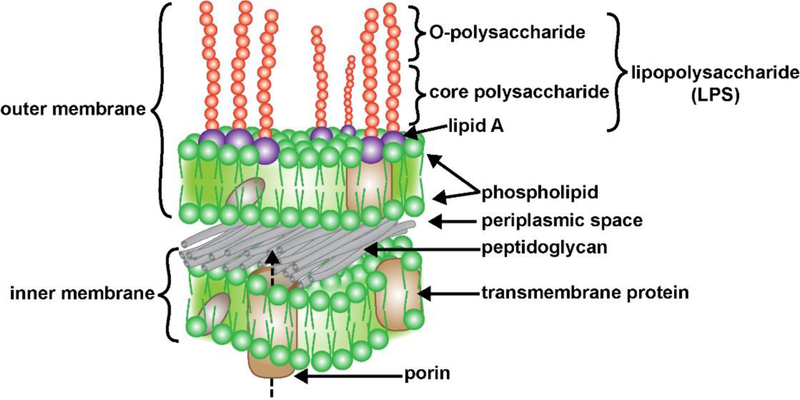
FIGURE 1 -.
Typical structure and organization of an anaerobic Gram-negative bacterial cell membrane and its containment of a lipopolysaccharide (LPS) coated surface (purple and red spheres); the two horizontal layers include an external (outer) and an internal (inner) membrane, both layers contain both integral (gray) and transmembrane (beige) globular proteins; the membranes are separated by an interwoven peptidoglycan layer and a periplasmic space; transmembrane protein complexes such as porin that transverse the inner membrane facilitate molecular communication and LPS transport between the bacterial cell interior to the bacterial outer membrane surface (dashed arrow) but the mechanisms are not well understood [20,47,57–59]. The outer surface of the external membrane contains a dense layer of LPS with lipids anchored in the membrane (purple spheres), and long core polysaccharide and O-polysaccharide side chains extending outward (red spheres); the externally facing LPS are highly thermostable, neurotoxic, pathogenic, extremely pro-inflammatory and a potent trigger of robust antigenic responses within the human immune system; LPS are constantly shed into the external environment where they may find their way past the GI-tract barrier into the systemic circulation and past the BBB into the brain parenchyma [1–5,9,15–18,39,50,57–62]; the mechanism of the induction of LPS and other GI-tract microbiome-derived neurotoxins by aluminum sulfate is not known; (source; figure adapted from https://www.dreamstime.com/stock-illustration-structure-gram-negative-bacteria-cell-wall-labeled-d-illustration-image84181743; last accessed 7 October 2019).

