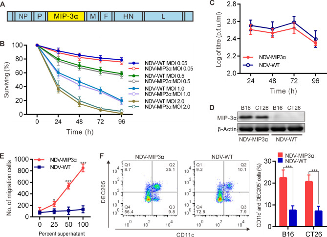
Figure 1.
Generation and evaluation of the recombinant virus NDV-MIP3α. (A) Schematic presentation of the recombinant NDV-MIP3α, showing the insertion site of the MIP-3α cDNA. (B) Lysis of B16 cells by NDV-MIP3α and NDV-WT at the indicated MOI and time points. (C) Virus replication of 1 MOI NDV-MIP3α or NDV-WT at the indicated time points in B16 cells. (D) MIP-3α expression in B16 and CT26 cells infected with 1 MOI NDV-MIP3α or NDV-WT. (E) Ex vivo chemotaxis for DCs by the supernatants from B16 cells infected with 1 MOI NDV-MIP3α or NDV-WT. (F) In vivo chemotaxis for DCs among the inflammation cells by FCM after the injection of the supernatants (100 µL at 48 hours) from B16 cells infected with 1 MOI NDV-MIP3α or NDV-WT. Data are plotted as mean±SD, *p<0.05, **p<0.01, ***p<0.001. DCs, dendritic cells; FCM, flow cytometry; MIP-3α, macrophage inflammatory protein-3α; MOI, multiplicity of infection; NDV, Newcastle disease virus; NDV-WT, wild-type NDV.

