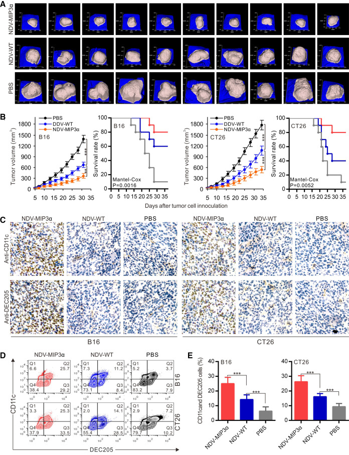
Figure 4.
NDV-MIP3α improves tumor control and enhances DC accumulation. B16 or CT26 tumor-bearing mice were intratumorally injected with NDV-MIP3α (2×10⁷ pfu) or NDV-WT (2×10⁷ pfu) or PBS after tumor cell inoculation. The tumor images and volumes were collected by a handheld device (TM900) in a 3-day interval. The tumor tissues were collected on day 25 after tumor cell inoculation. (A) The representative images of B16 tumor masses on day 28 after tumor cell inoculation. (B) Data of the tumor volumes and survival rates of the tumor-bearing mice at the indicated time points in B16 and CT26 tumor-bearing mice (n=10). (C) The representative section images from the indicated tumor masses stained with anti-CD11c and anti-DEC205 antibodies. (D) The representative images of CD11c and DEC205 double-positive TILs detected by FCM and (E) data of three independent experiments. Data are plotted as mean±SD, **p<0.01, ***p<0.001. FCM, flow cytometry; MIP-3α, macrophage inflammatory protein-3α; MOI, multiplicity of infection; NDV, Newcastle disease virus; NDV-WT, wild-type NDV; TILs, tumor-infiltrating lymphocytes.

