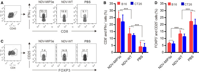
Figure 7.
NDV-MIP3α modulates the tumor microenvironments. The TILs were isolated from the tumor masses of B16-bearing and CT26-bearing mice, CD45 or CD4 cells were gated as indicated and analyzed by FCM. (A) The representative images of FCM-analyzed CD45+ and CD8+ lymphocytes secreting IFN-γ in the tumor-bearing mice treated with the indicated NDV or PBS. (B) The data of four independent experiments of flow cytometry (FCM) show the percentage of CD45+ and CD8+ lymphocytes secreting IFN-γ. (C) Representative images of FCM-analyzed CD4+, CD25+, and Foxp3+ triple-positive Tregs in the mice treated with the indicated NDV or PBS. (D) The data of four independent experiments of FCM show the percentage of CD4+, CD25+, and Foxp3+ triple-positive Tregs. Data are plotted as mean±SD, ***p<0.001. FCM, flow cytometry; MIP-3α, macrophage inflammatory protein-3α; NDV, Newcastle disease virus; NDV-WT, wild-type NDV; TILs, tumor-infiltrating lymphocytes; Tregs, regulatory T cells.

