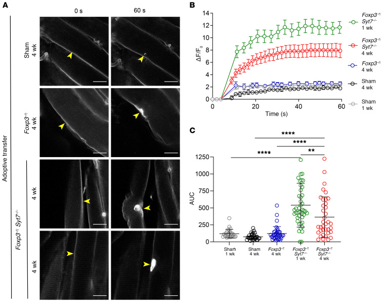
Figure 4. Sarcolemmal resealing capacity is significantly diminished following adoptive transfer of lymph node cells from Foxp3–/Y Syt7–/– mice.
FDB muscles isolated from indicated mice and injured with an infrared laser in the presence of FM4-64 dye. The kinetics of sarcolemma resealing was measured by acquisition of images every 3 seconds for 60 seconds and calculation of the change in fluorescence before and after injury. (A) Representative images of Rag1–/– mice receiving sham adoptive transfer, adoptive transfer of Foxp3–/Y lymph node cells (4 weeks), or adoptive transfer of Foxp3–/Y Syt7–/– lymph node cells (1 week and 4 weeks). Scale bars: 20 μm. Yellow arrowheads indicate sites of injury. (B) Curves depicting mean sarcolemmal resealing kinetics measured every 3 seconds for 60 seconds. Data are represented as mean ± SEM. (C) AUC calculations representing total dye influx over time (Sham 1 week, n = 27; Sham 4 weeks, n = 31; Foxp3–/Y adoptive transfer, n = 37; Foxp3–/Y Syt7–/– adoptive transfer 1 week and 4 weeks, n = 44 and 37, respectively; ANOVA, F(4,171) = 28.47, P < 0.0001; Tukey’s HSD: Sham 1 week vs. Foxp3–/Y Syt7–/– 1 week, P < 0.0001; Sham 4 weeks vs. Foxp3–/Y Syt7–/– 4 weeks, P < 0.0001; Foxp3–/Y 4 weeks vs. Foxp3–/Y Syt7–/– 4 weeks, P = 0.0005). Data are represented as mean ± SD.

