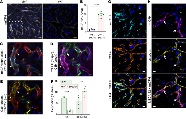
Figure 7. Recombinant factor H binds in the sinusoids of fH–/– mice.
(A and B) rmCFH binds within the sinusoids of fH–/– mice in a continuous pattern (A, left), but is reduced and punctate in WT mice (A, right). Scale bars: 20 μm. (B) Quantification of bound rmCFH. ****P < 0.0001, t = 12.98, df = 4.544; 2-tailed t test with Welch’s correction; mean ± SEM. For A and B, n = 5 males per group (WT, 9 months; fH–/–, 7 months). (C–E) Enlarged view of fH–/– image identified by dashed white box in A. Abundant rmCFH deposition (arrowhead) near iC3b/C3d (C, turquoise) and C3b (D, red). Intermittent C3b (C and E; arrows) colocalized with linear iC3b/C3d. Scale bars: 10 μm. (F) Compared with unmanipulated fH–/–, rmCFH-reconstituted fH–/– mice have reduced sinusoidal C3b (****P < 0.0001, t = 8.424, df = 7.179) and increased iC3b/C3d, demonstrating rmCFH-mediated conversion of C3b to iC3b/C3d (**P = 0.0015, t = 4.729, df = 7.931; 2-tailed t test with Welch’s correction; mean ± SEM; n = 5 males [7 months] per group). (G and H) rmCFH colocalized with COL4 (G) and MECA-32 (H, arrows) in fH–/– sinusoids, but not on the large vasculature (vascular lumen denoted by asterisks). Nuclei stained with DAPI (blue). Scale bars: 10 μm; n = 5. Representative images shown from 4 independent experiments.

