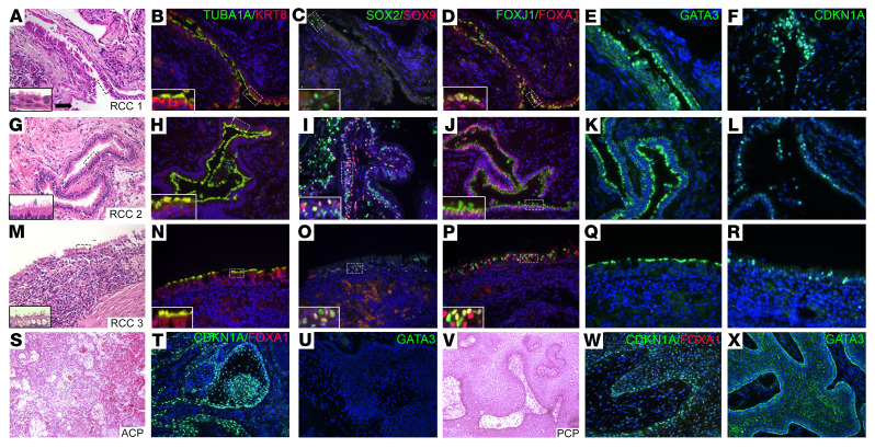
Figure 5. hRCCs express FOXA1.
(A–R) Classification of hRCC samples (n = 16) was confirmed by staining with H&E to detect ciliated cells (insets) (A, G, and M) and costaining with antibodies for cytokeratin8 and acetylated tubulin (B, H, and N). Costaining with antibodies for progenitor markers SOX2 and SOX9 detected many positive cells (C, I, and O). Costaining with antibodies FOXJ1 and FOXA1 detected positive cells lining the cysts (D, J, and P). Immunostaining for GATA3 (E, K, and Q) and CDKN1A (F, L, and R) detected positive cells lining the cysts in some, but not all samples. (S–X) Surgical samples from craniopharyngiomas of both the ACP (n = 7) (S–U) and PCP (n = 5) (V–X) subtypes were stained with H&E, costained for CDKN1A and FOXA1, and immunostained for GATA3. Samples were positive for CDKN1A and negative for both FOXA1 and GATA3. Scale bar: 50 μm.

