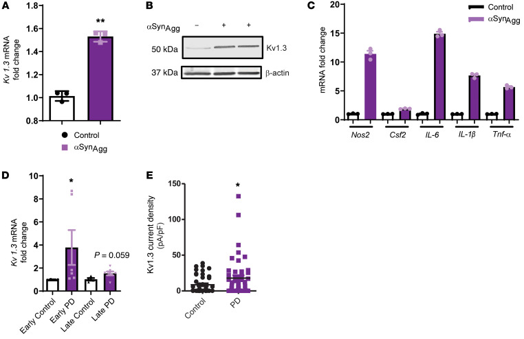
Figure 2. Upregulated expression of the potassium channel Kv1.3 upon aggregated αSyn stimulation in ex vivo slices and B cells derived from patients with PD.
(A) Midbrain slice cultures were treated with 1 μM αSynAgg for 24 hours. qRT-PCR shows upregulated Kv1.3 mRNA expression. (B) Western blot shows upregulated Kv1.3 protein level in midbrain slice cultures treated with 1 μM αSynAgg for 24 hours. (C) qRT-PCR of midbrain slice cultures treated with 1 μM αSynAgg for 24 hours, revealing upregulation of the proinflammatory factors Nos2, Csf2, IL-6, IL-1β, and Tnfa. (D) qRT-PCR shows increased Kv1.3 mRNA expression in B cell lymphocytes isolated from patients with PD compared with expression in B cell lymphocytes from age-matched controls. (E) Whole-cell patch clamping of B cell lymphocytes isolated from patients with PD showed higher Kv1.3 channel activity compared with that observed in age-matched controls (n = 3 control and n = 3 PD). A 1-way ANOVA was used to compare multiple groups in C and D. Tukey’s post hoc analysis was applied. A 2-tailed Student’s t test was used to compare 2 groups. Each dot on the bar graphs represents a biological replicate. Data are presented as the mean ± SEM, with 3–7 biological replicates from 2–3 independent experiments unless otherwise indicated. *P ≤ 0.05 and **P < 0.01.

