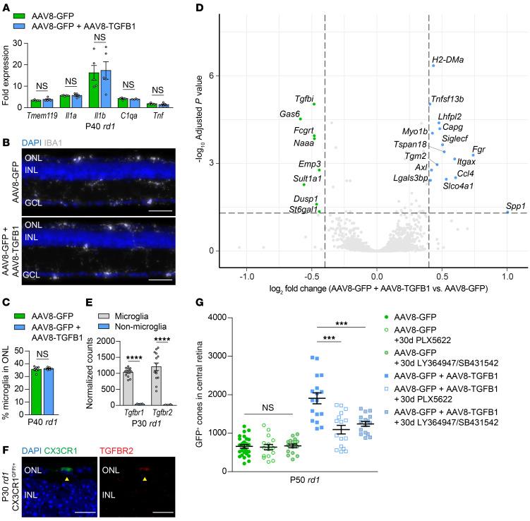
Figure 4. Role of retinal microglia in AAV8-TGFB1-mediated cone survival.
(A) mRNA expression of indicated genes in FVB (rd1) retinas (n = 4–5) treated with AAV8-GFP or AAV8-GFP plus AAV8-TGFB1. Fold changes are relative to untreated WT (sighted FVB) retinas. Representative images (B) and quantification (C) of IBA1-positive microglia in the ONL of P40 rd1 retinas (n = 6–7) treated with AAV8-GFP or AAV8-GFP plus AAV8-TGFB1. Scale bar: 50 μm. (D) Volcano plot of up- and downregulated genes in microglia sorted from P30 rd1 retinas (n = 7) after treatment with AAV8-GFP plus AAV8-TGFB1 relative to AAV8-GFP only. Dotted lines indicate adjusted P < 0.05 and log2 fold change >0.4. (E) Normalized RNA-seq counts for expression of Tgfbr1 and Tgfbr2 in microglia versus nonmicroglia cells from P30 rd1 retinas (n = 4–14). (F) Immunostaining for TGFBR2 in rd1 CX3CR1GFP/+ retinas (n = 2). Arrowheads indicate colocalization of TGFBR2 with a CX3CR1-positive microglia in the ONL. Scale bar: 10 μm. (G) Quantification of GFP-positive cones in central retinas of rd1 mice after 30 days of microglia depletion with PLX5622 (n = 16) or inhibition of TGFBR1/2 with LY364947 and SB431542 (n = 16). Data for untreated groups are taken from Figure 2E. Data shown are mean ± SEM. ***P < 0.001, ****P < 0.0001 by 2-tailed Student’s t test with Bonferroni’s correction for A and G, 2-tailed Student’s t test for C and E. INL, inner nuclear layer.

