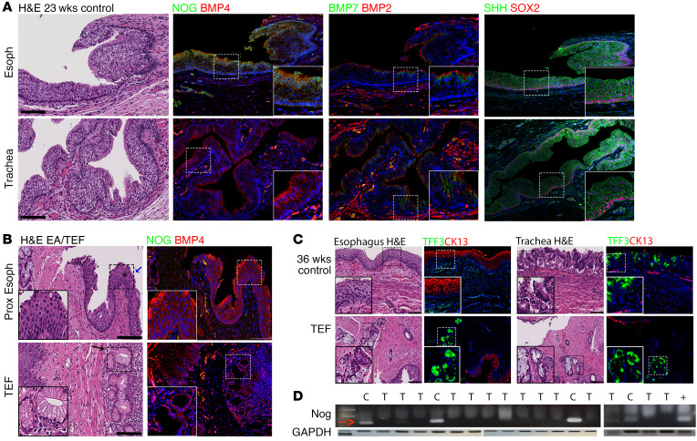
Figure 1. NOG is selectively expressed in developing human esophagus but not in the trachea, and EA/TEF tissue lacks NOG.
(A) Pathology samples from human autopsies stained for genes that have been previously known to be important in foregut development. Paraffin sections were used to perform IHC with NOG, BMP7, SHH (green), BMP4, BMP2, and SOX2 (red). All genes were present in both esophagus and trachea. However, NOG was missing from respiratory tissue. (B) Pathology samples from EA/TEF repair were collected to study NOG and BMP expression using IHC. On H&E staining, a combination of esophageal and respiratory glands were found in atretic esophagus as well as in fistula. NOG is absent from EA/TEF samples when evaluated by IHC as well as RT-PCR (B, D). (C) Human esophageal and respiratory pathology samples stained for CK13 (red) and TFF3 (green). TEF from human neonate expresses both CK13 and TFF3. (D) RNA was isolated from paraffin samples of either esophagus of control neonates, or EA/TEF tissue obtained at the time of surgical repair. Positive control (+) is RNA extracted from Kato III cell line. *Pseudostratified columnar epithelium with cilia. Blue arrow indicates cilia. Black arrow indicates seromucinous glands. Inserts are ×2 magnification. Scale bars: 100 μm. C, control, T, TEF.

