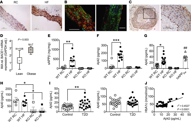
Figure 1. BACE1 vascular expression and activity and plasma levels of Aβ in mice and humans.
(A) Immunohistochemical staining for BACE1 (brown) showing representative sections from aortas of RC-fed and HF-fed (DIO) WT mice. (B) Confocal images of DIO mouse aortas stained for BACE1 (green) and SMA (left) or CD31 (right), respectively (red). Scale bars: 50 μm. (C) Immunohistochemical staining for BACE1 in human nonatherosclerotic temporal artery, with a section (rectangle) at higher magnification. (D) BACE1 mRNA expression in internal mammary arteries from lean and obese individuals (Mann-Whitney U test). HF feeding increases BACE1 activity in WT mouse aorta, as determined by sAPPβ (E) and Aβ42 (F) levels, with BACE1-KO aortas showing negligible peptide levels (n = 4–12). (G) Plasma Aβ42 levels are increased in DIO mice to RC-fed hAPPSw mouse levels, with insignificant plasma Aβ42 in RC- or HF-fed BACE1-KO mice (n = 6–14). (H) Plasma Aβ40 levels of RC-fed and DIO mice and RC- and HF-fed BACE1-KO mice (n = 6–14). (I) Plasma Aβ42 and Aβ40 levels in control (n = 20) and obese individuals with type 2 diabetes (T2D) (n = 20). (J) Linear regression between plasma Aβ42 and HbA1c in control and obese individuals with T2D (P < 0.001). Data presented as means ± SEM for all figures except D, where SD given. *P < 0.05; **P < 0.01; ##P < 0.01; ***P < 0.001 by 2-way ANOVA with Tukey’s multiple-comparisons test (E–H) or 2-tailed unpaired Student’s t test (G and I).

