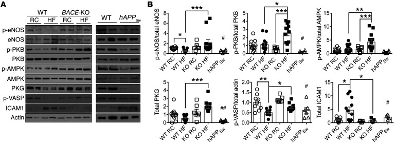
Figure 6. Aortic NO signaling is modified in BACE1-KO and hAPPSw mice.
(A) Representative immunoblots of p-eNOS(Ser1177), eNOS, p-PKB(Ser473), PKB, p-AMPK(Thr172), AMPK, PKG, p-VASP(Ser239), ICAM1, and actin in aortas of RC-fed WT, DIO, and RC- and HF-fed BACE1-KO mice (left panel), and from aortas of RC-fed WT and RC-fed hAPPSw mice (right panel). (B) Ratio of signal intensities for p-eNOS to eNOS, p-PKB to PKB, p-AMPK to AMPK; and for p-VASP, PKG, and ICAM1 to actin (n = 6–15). Data are means ± SEM. *P < 0.05; **P < 0.01; ***P < 0.001 by Kruskal-Wallis test with Dunn’s multiple-comparisons test. #P < 0.05, ##P < 0.01 by Mann-Whitney U test (vs. RC-fed WT mice).

