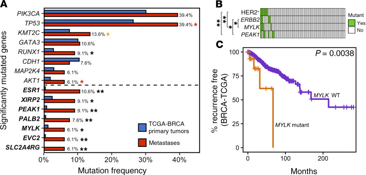
Figure 2. Genes preferentially mutated in metastases.
(A) Mutation frequencies for SMGs identified by MutSigCV2 within metastatic tumors in our cohort (red, n = 66) and primary tumors in TCGA-BRCA (blue, n = 1044). Seven SMGs, indicated in bold, have not been reported in TCGA-BRCA primary tumors across nor within subtypes. Eleven SMGs exhibited significantly higher mutation frequencies in metastases within our cohort compared with TCGA-BRCA primary tumors (2-sided Fisher’s exact test; *FDR ≤ 0.10; **FDR < 0.001). Red and orange asterisks respectively denote 3 SMGs that either lose or gain significance when less stringent filtering criteria employed in Ciriello et al. (3) are used. (B) Co-occurrence of MYLK and PEAK1 mutations with ERBB2 mutations and HER2+ status in metastases (2-sided Fisher’s exact test, *P < 0.05; **P < 0.01; FDRs = 0.07–0.36). Each column represents a metastatic tumor. (C) Kaplan-Meier survival analysis showing that TCGA-BRCA patients whose primary tumor had a mutation in MYLK exhibited shorter RFS.

