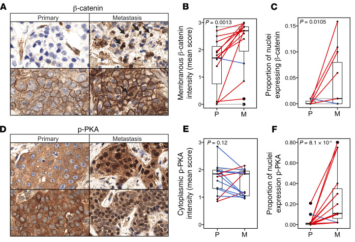
Figure 7. Preferential WNT activation and nuclear localization of activated PKA in metastases.
IHC analyses of β-catenin (A–C) and p-PKA (D–F) in primary (P) and metastatic (M) tumors (n = 15). (A) Representative β-catenin IHC images for primary and metastatic tumors. (B) Mean membranous β-catenin staining intensity and (C) proportion of nuclei that are β-catenin–positive in paired and matched primary and metastatic tumors (1-sided Wilcoxon’s signed-rank test). (D) Representative p-PKA IHC images for primary and metastatic tumors. (E) Mean cytoplasmic p-PKA staining intensity and (F) proportion of nuclei that are p-PKA–positive in paired and matched primary and metastatic tumors.

