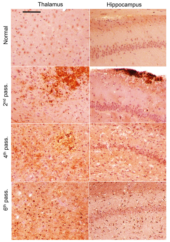
Figure 2. Change in PrPSc deposition upon serial passaging.

Representative images of PrPSc deposition in the thalamus and hippocampus of animals from passages 2, 4, and 6 of SSLOW-Mo and normal controls. Antibody SAF-84 was used for staining. Scale bar: 100 μm. Brains of normal-age mice (337–405 days old) were used for reference.
