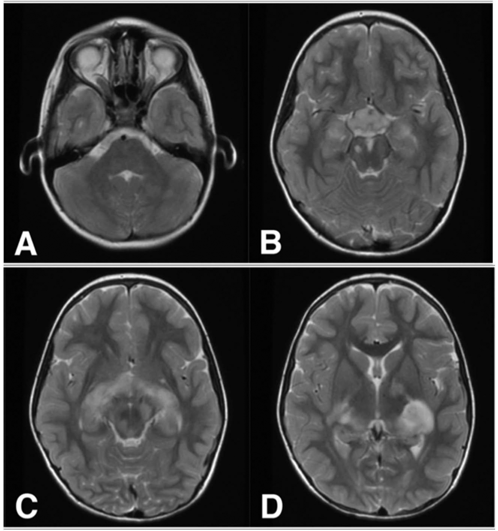
FIG. 1.

A 7-year-old girl with NF1-OPG involving the optic nerves (A), optic chiasm and hypothalamus (B), and optic tracts (C) shown on T2 sequences. Focal areas of T2 signal intensity are demonstrated in the midbrain and thalamus (B–D) and may abut tumor margins. NF1, neurofibromatosis type 1; OPG, optic pathway glioma.
