Fig. 5.
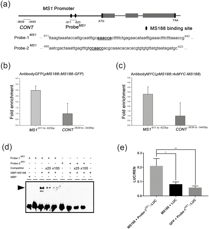
MS188 directly regulates MS1 in vivo and in vitro. (a) Potential MYB-binding sites in the promoter and genome regions of the MS1 gene. Black ellipses indicate the potential MS188-binding sites. ProbeMS1 contains these binding sites. (b, c) The enrichment of the MS1 promoter was confirmed by ChIP-qPCR with primer sets [MS1−911 to −623 bp, CONTMS1(−3639 to −3445 bp)] using the pMS188:MS188-GFP (b) and the pMS188:4×MYC-MS188 (c) samples. The fold enrichment was calculated from three independent replicates. Error bars represent the SD (n=3). (d) ProbeMS1 was divided into two segments containing the MYB-binding site named Probe-1MS1 (MS1−911 to −842 bp) and Probe-2MS1 (MS1−685 to −623 bp). MBP–MS188 protein was mixed with a biotin-labelled probe, a 25-fold and 100-fold unlabelled competitor probe for EMSA assay. The arrowhead indicates a band shift. (e) Dual-LUC assay measuring the MS188 transactivation on the Probe-1MS1-driven LUC reporter. The MS188 effector (AtUbi10:MS188) paired with empty LUC reporter and free GFP (AtUbi10:GFP) paired with the Probe-1MS1-driven LUC reporter were co-transfected to serve as controls. One-way ANOVA and pairwise Dunnett test were used to test the statistical difference between groups (*P<0.05, **P <0.01). Error bars stand for Probe-1 SD.

