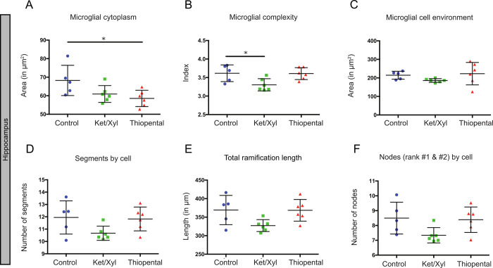
Fig 4. Hippocampus variability by morphological criteria in the different conditions.
Microglial morphology was characterized using the following parameters: microglial cytoplasm (A), the complexity index (B), the cell environment area in μm2 (C), the number of segments by cell (D), the total ramification length in μm (E) and the number of nodes (rank #1 & #2) by cell (F). Data shown are mean±SD in the control, ketamine/xylazine and thiopental conditions (n = 5, n = 6 and n = 6, respectively). In CX3CR1GFP/+ mice, 272 to 448 microglial cells were analyzed by region and by animal, resulting in studying respectively n = 1713, 1808 and 2173 cells by condition. ANOVA Kruskal-Wallis test was used to compare the different regions. *p<0.05.

