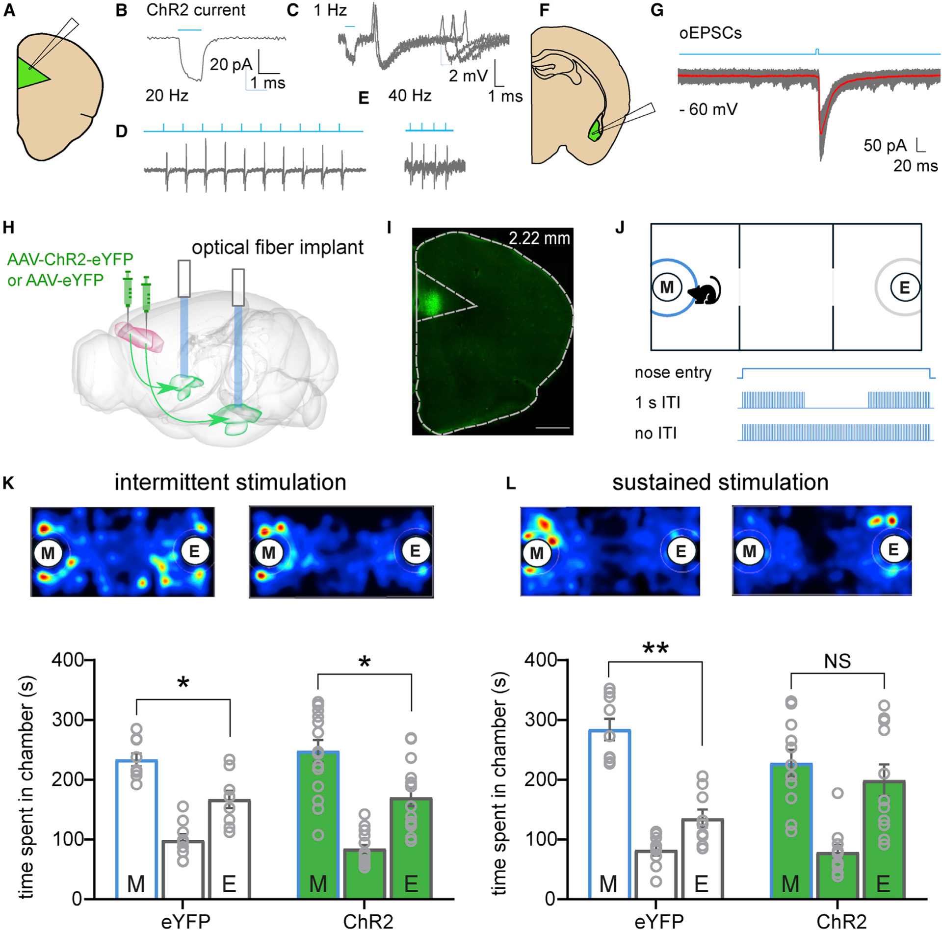
Figure 2. Temporally Cued Activation of PL-to-BLA Circuitry during Social Investigation Impairs Social Behavior.

(A) Cartoon of recording configuration in the PL.
(B) Voltage-clamp recording showing depolarized membrane potential during the presence of blue light.
(C–E) Blue light-evoked action potentials recorded from putative pyramidal cells at 1 Hz (C), 20 Hz (D), and 40 Hz (E).
(F) Cartoon of recording configuration in the BLA.
(G) Optically evoked excitatory postsynaptic current (oEPSC) from putative principal neuron.
(H) Diagram showing injection of AAV-ChR2-eYFP or control virus AAV-eYFP in the PL and optical fiber ferrule implants targeting the BLA.
(I) Epifiuorescence image of injection site confirming transduction in PL. Green is ChR2-eYFP. Scale bar: 500 μm.
(J) Schematic of optogenetic three-chamber apparatus. Investigation zone in blue depicts area that triggers light stimulation upon mouse nose entry.
(K and L) Representative heatmaps and quantification of time spent in each chamber.
(K) Intermittent light stimulation during social investigation did not affect social preference in control or ChR2 groups. n = 9 for control mice and n = 15 for ChR2-expressing mice.
(L) Sustained light stimulation during social investigation abolished the social preference in ChR2-expressing mice but not in controls. n = 9 for control mice and n = 11 for ChR2-expressing mice. Paired-samples t tests were used to compare time spent in the chambers containing a mouse in a tube and an empty tube separately for each group in (K) and (L).
Data are represented as mean ± SEM. *p < 0.05. See also Figure S2 relating to these experiments.
