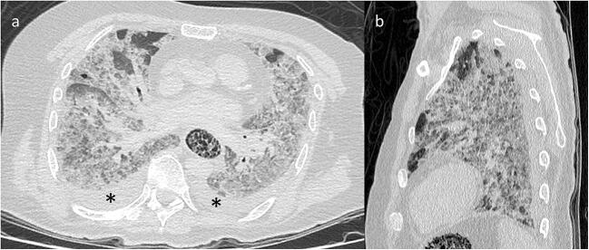
Fig. 1.
Post-mortem computed tomography and pulmonary parenchyma reconstruction. Axial (a) and sagittal (b) views showing diffuse, bilateral and panlobar ground-glass opacities associated with interlobular and intralobular septal thickening, subpleural consolidations and bilateral pleural effusions (asterisk). The anterior portions of both lungs were more likely to be spared

