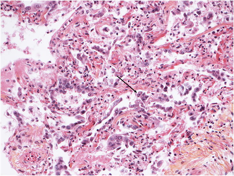
Fig. 3.

Microscopic findings in the lungs. (HES, magnification × 20). Diffuse alveolar damage in a more organized stage. Note the enlargement of the alveolar septa, fibrin deposition and incorporation, intraalveolar exudates and a type-2 pneumocyte hyperplasia (arrow) representing patchy, interstitial chronic inflammation
