
Extended Data Figure 8: Immunohistochemical staining of fibrillin-1.
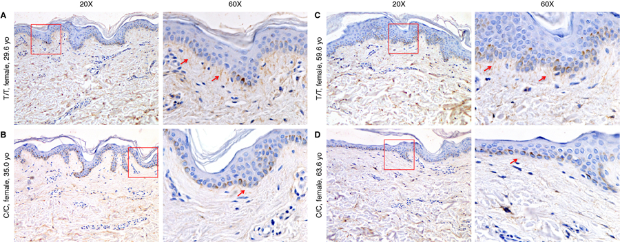
A–B) Fibrillin-1 staining in skin biopsies in two individuals with rs200342067 C/C genotype and C–D) two individuals with T/T genotype matched for age, sex, and ancestry proportions. Individuals with C/C genotype have less fibrillin-1 deposition in the dermal extracellular matrix (ECM) and shorter microfibrillar projections from the dermal-epidermal junction into the superficial (papillary) dermis (red arrows, 20x) as well as less fibrillin-1 deposition in the deeper dermis. Two magnification have been shown, the red rectangles in the first column (20x magnification) are magnified in the second column (60x).