
Figure. 6.
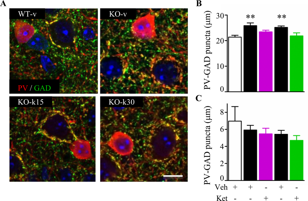
Prolonged ketamine treatment restores parvalbumin-circuit inputs onto pyramidal cells. (A) Representative confocal high magnification images showing parvalbumin (PV, green) and GAD65 (GAD, red) in WT-v, KO-v, KO-k15 and KO-k30. Scale bar 10 mm. (B) PV-cell innervations of pyramidal cell somata were statistically increased in KO-v compared to WT-v (Kruskal-Wallis, ** p ≤ 0.01, Dunn’s post-test). Both ketamine treatments reduced PV-innervations towards WT levels. (C) PV-PV connections were not affected by the loss of Mecp2 or by the ketamine treatments. (WT-v15, n = 3; KO-v15, n = 3; KO-k15, n = 4; WT-v30, n = 3; KO-v30, n = 4; KO-k30, n = 4 mice). Data are expressed as mean ± SEM.