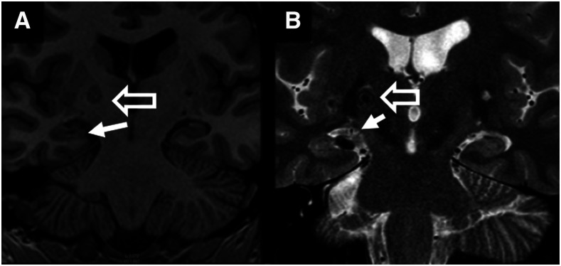
Figure 4.
Hippocampal sclerosis. Hippocampal sclerosis (defined as the presence of hippocampal atrophy and a hyperintense signal on long-repetition-time sequences of the hippocampus) is seen on the R-side (solid arrow) of the T1 (A) and T2-FLAIR (B) coronal MRI scans. An adjacent calcified lesion (hollow arrow) is also shown.

