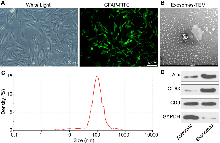
Figure 1.
Identification of ATCs and exosomes. (A) Morphological analysis and immunofluorescence staining of ATC-specific marker GFAP showed that the cells we used were ATCs. (B) ATCs-derived exosomes were elliptical- or cup-shaped with a size of about 100 nm observed under a Transmission electron microscopy. (C) Nanosight nanoparticle tracking analysis indicated that the particle size was about 100 nm. (D) Exosome marker proteins CD9, CD63 and Alix determined by RT-qPCR. All experiments were performed three individual times.
Abbreviations: ATC, astrocyte; GAPDH, glyceraldehyde-3-phosphate dehydrogenase; GFAP-FITC, glial fibrillary acidic protein-fluorescein isothiocyanate.

