Figure 2.
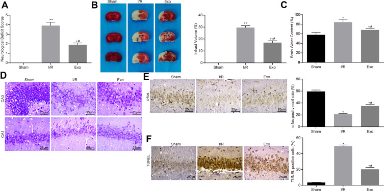
ATC-derived exosomes reduce I/R-induced neurological damage in rats. (A) After the injection of 30 μg/mL Exo in the I/R group, the neurological deficit score was used to evaluate the effects of ATC-derived exosomes on neurocognitive function in rats. (B) TTC staining was used to calculate the cerebral infarct size in rats treated with exosomes. (C) The degree of cerebral edema was detected in rats treated with exosomes. (D) Nissl staining was used to detect the number of Nissl’s body in rat brain tissue. (E) Detection of nerve activity in rats in each group by c-fos immunohistochemistry. (F) Detection of neuronal apoptosis in rats in each group by TUNEL. All experiments were performed three individual times; Data are expressed as mean ± standard deviation. One-way ANOVA and Tukey’s multiple comparison test were used to determine statistical significance. *P < 0.05, **P < 0.01 vs the Sham group; #P < 0.05 vs the I/R group.
Abbreviations: ANOVA, analysis of variance; ATC, astrocyte; Exo, exosome; I/R, ischemic-reperfusion; TTC, 2,3,5-triphenyltetrazole chloride; TUNEL, terminal deoxynucleotidyl transferase (TdT)-mediated dUTP nick end labeling.

