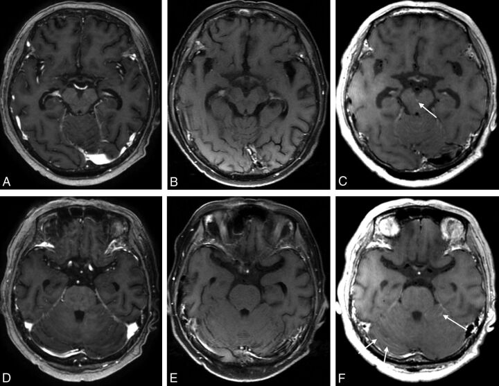
Fig 2.
MR images of a 60-year-old female patient with lung cancer. The contrast-enhanced 3D ultrafast GRE (A and D) and SE T1WI (B and E) were negatively interpreted by all raters. On only the black-blood imaging (C and F) scans was leptomeningeal enhancement along the interpeduncular cistern (C, arrow) and bilateral cerebellar surface observed (F, arrows).

