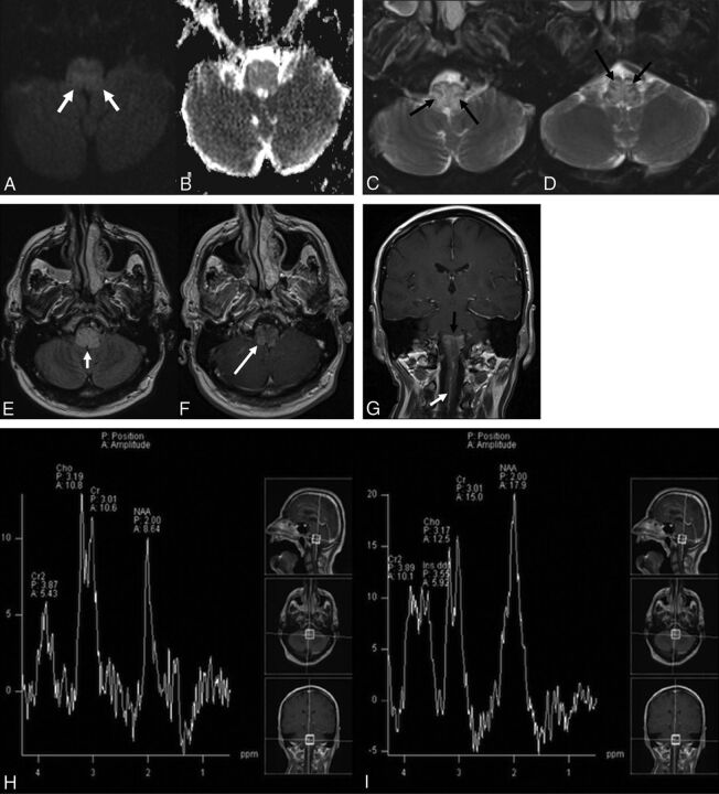
Fig 1.
A 59-year-old man who presented with new-onset dizziness and severe nausea and vomiting. Initial MR imaging demonstrates mild hyperintense signal on axial DWI (b = 1000) (white arrows, A) without definite restricted diffusion on the corresponding ADC map (B). There is relatively diffuse hyperintense signal abnormality within the medulla on axial T2WI with fat suppression (C and D) with sparing of the periphery and several internal linear regions (black arrows). This pattern of sparing was not initially recognized. E, Axial FLAIR imaging demonstrates relatively diffuse hyperintense signal abnormality within the medulla (short white arrow). Axial postcontrast T1WI (F) demonstrates mild patchy enhancement (long white arrow). G, Coronal postcontrast T1WI reveals a dilated perimedullary vein (white arrow) extending inferiorly from the patchy enhancement within the medulla (black arrow) and coursing caudally along the upper cervical cord. MR spectroscopy with TEs of 135 (H) and 35 (I) ms demonstrates no significant elevation in choline with decreased NAA. MR perfusion imaging (J) reveals slightly decreased relative CBV within the medulla. Suspicion was raised for an underlying DAVF. Left external carotid injection on digital subtraction angiography arterial phase lateral (K) and delayed venous phase anteroposterior (L) views demonstrates a DAVF in the wall of the left superior petrosal sinus (long white arrow) fed by meningeal branches of the occipital (short white arrow), ascending pharyngeal (long black arrow), and middle meningeal (short black arrows) arteries, with retrograde venous drainage via the petrosal vein to the pial perimedullary veins running to the cervical cord as the anterior and posterior spinal veins (black dashed arrows).


