Fig 3.
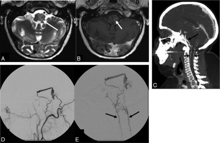
A 35-year-old woman with a history of pilocytic astrocytoma as a child now presenting with 4 weeks of progressive difficulty in walking. A, Axial T2WI reveals patchy hyperintensity within the medulla (long white arrow) with peripheral sparing. B, Axial postcontrast T1WI demonstrates subtle intramedullary enhancement (short white arrow). C, Sagittal CTA reformatted image depicts prominent perimedullary (long black arrow) as well as ventral and dorsal spinal veins (short black arrows). Selective DSA of the occipital (D) and mastoid branch via microcatheter (E) demonstrates the fistulous connection in the wall of the superior petrosal sinus with retrograde pial reflux via the petrosal vein to the perimedullary and spinal veins (black arrows).

