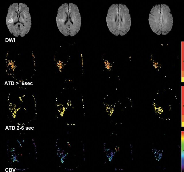
Fig 2.
A 70-year-old woman with left paresis who had right MCA (M1) occlusion (not shown) and insufficient collaterals on baseline conventional angiography. DWI showed right MCA territorial infarction. Processed perfusion maps show 17 mL of severe (ATD>6 seconds) hypoperfusion, 20 mL of moderate (ATD2–6 seconds) hypoperfusion, and a mean rCBV2–6 seconds of 0.9 within the hypoperfused area. The perfusion collateral index is 20 × 0.9 = 18.

