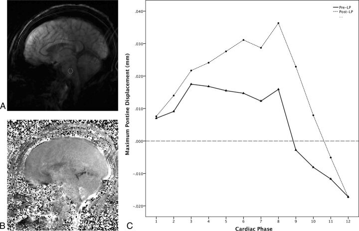
Fig 1.
DENSE imaging with ROI placement and measured displacement across the cardiac cycle from a 44-year-old woman with IIH (Patient 9). A, Sagittal magnitude image from pre-LP DENSE encoded for motion in the foot-to-head direction. An ROI has been placed in the midpons to avoid partial volume effects from CSF flow. B, Corresponding phase image from the pre-LP DENSE encoded for motion in the foot-to-head direction with ROI transferred from the magnitude image into the midpons for measurement to be propagated to all 12 images acquired across the cardiac cycle. C, Graph showing DENSE displacement in the foot-to-head direction across the cardiac cycle divided into 12 phases. The solid line shows the pre-LP displacements across the cardiac cycle, and the dotted line shows post-LP displacements across the cardiac cycle. Maximum displacement is calculated by subtracting the lowest value from the highest across the cardiac cycle for both the pre-LP and post-LP states. Note the increased displacement in the post-LP state.

