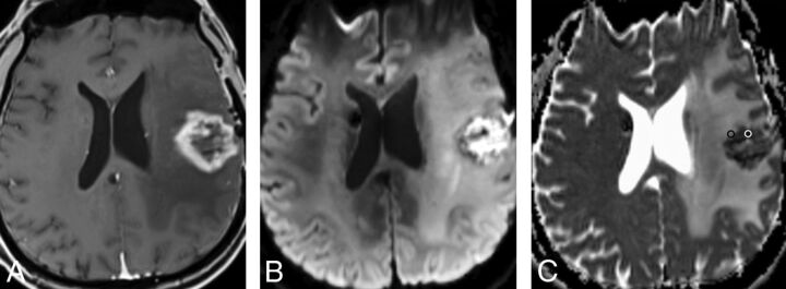
Fig 1.
Radiation necrosis: A, Axial T1-weighted MR image shows a left frontal necrotic ring-enhancing lesion. B and C, Axial DWI and ADC map demonstrate diffusion restriction within the necrotic center of the lesion. Representative ROIs are placed on the ADC map corresponding to the enhancing rim (black ROI) and the necrotic component (white ROI).

