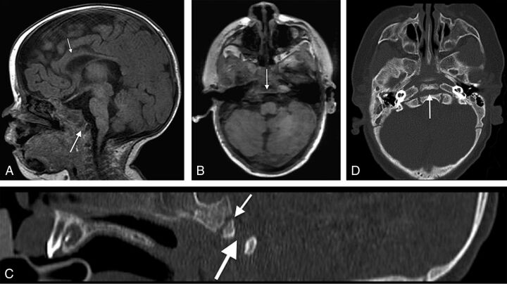
Fig 2.
A, Midline sagittal spoiled gradient-recalled acquisition T1WI (1.5T MR imaging; TR/TE = 12/5 ms; TI = 500 ms; slice thickness = 2 mm) from a 4-month-old girl with CHARGE syndrome shows a complete coronally oriented clival cleft in the basiocciput (large arrow). Also note thinning of the corpus callosum (small arrow), cerebellar vermian hypoplasia, and brain stem volume loss. B, Axial spoiled gradient-recalled acquisition of the same patient shows a coronally oriented cleft traversing the clivus (arrow). Corresponding sagittal (C) and axial (D) CT images demonstrate the complete cleft (large arrow) as distinct from the normal spheno-occipital synchondrosis (small arrow in C).

