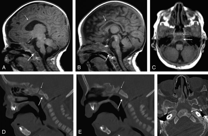
Fig 3.
Spoiled gradient-recalled acquisition T1WI (3T MR imaging; TR/TE = 6.6/2.5 ms; TI = 700 ms; slice thickness = 0.63 mm) from a 34-day-old boy with CHARGE syndrome shows a partial (unilateral left side) coronally oriented clival cleft in the basiocciput. A, Left parasagittal unilateral clival cleft (large arrow), mild thinning of the corpus callosum (small arrow), cerebellar vermian hypoplasia, and brain stem volume loss are illustrated. B, Right parasagittal image shows a normal appearance of the clivus without clefting, a thin corpus callosum (small arrow), and vermian and brain stem hypoplasia. C, Axial T1WI of the same patient with a left-sided cleft (large arrow) and normal spheno-occipital synchondrosis (small arrow). Corresponding sagittal and axial CT images (D and F) further demonstrate the unilateral left clival cleft (large arrow, D and F) and absence of clival clefting on the contralateral side (E). Note normal spheno-occipital synchondrosis (small arrow, D and E).

