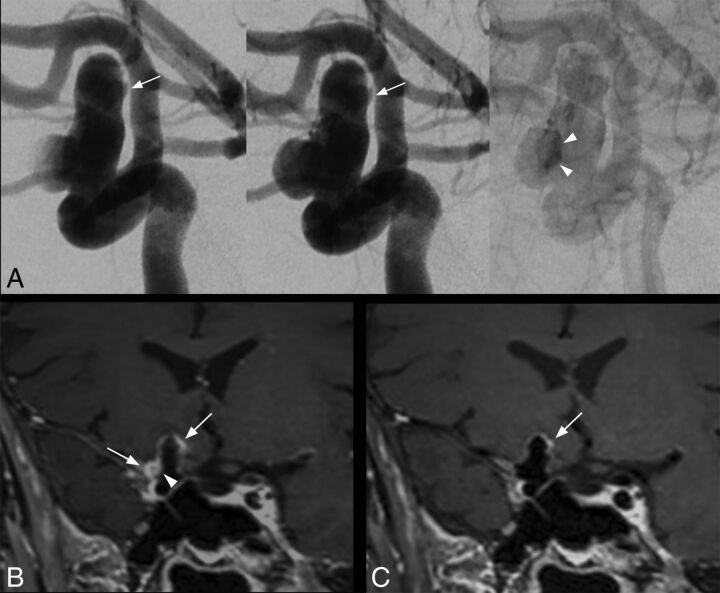
Fig 1.
Saccular intracranial aneurysm in a 63-year-old patient. DSA angiogram (A, from left to right: early arterial phase, arterial phase, and venous phase) shows a 14-mm right internal carotid artery aneurysm with intrasaccular slow flowing/stagnant blood (arrowheads) and irregular shape (arrow). With conventional SPACE imaging (B), extensive AWE is visible, more prominent at the apex and at the lateral portion of the aneurysm (B, arrows). Note the enhancement within the aneurysm lumen visible only using conventional SPACE imaging (B, arrowhead), matching the stagnant blood on DSA (A, arrowheads). With MSDE SPACE imaging, AWE is only visible at the apex (C, arrow).

