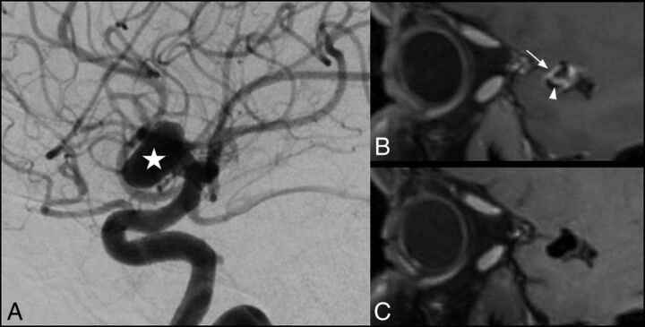
Fig 2.
Saccular intracranial aneurysm in a 42-year-old patient. DSA angiogram shows an 11-mm aneurysm (A, star) arising from the M1 segment of the right middle cerebral artery. AWE is visible on the conventional SPACE image (B, arrow), while there is no enhancement visible on the corresponding MSDE SPACE image (C). Enhancement within the aneurysm lumen was also visible using a conventional SPACE image (B, arrowhead).

