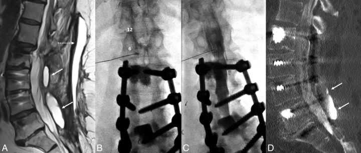
Fig 1.
A 67-year-old woman with a history of multiple spinal fusions presented with newly worsening low back and radicular leg pain (case 1). Sagittal T2-weighted MR imaging (A) shows 2 paraspinous fluid collections (white arrows) within the deep paraspinal musculature and a midline subcutaneous fluid collection (dashed arrow) with rim enhancement on postcontrast series (not shown). B, Anteroposterior fluoroscopy image of myelography with TFLP. The needle tip is beyond the medial edge of the pedicle at the 5 o'clock position (relative to the pedicle). Note the position of the needle inferior to the expected location of the exiting nerve root and dorsal root ganglion. C, Oblique fluoroscopy image shows contrast extending into the intrathecal space after injection through the left L1–2 foramen. D, CT myelogram demonstrates the inferiorly located cystic collection filled with contrast, confirming a pseudomeningocele (arrows).

